Charge coupled device detector (CCD) and solid state laser (DPSS) at 532 nm have been supplied for the first time after my arrival at Wakayama University ten years ago. This enables me to do research Raman spectrosopic measurements on condensed matters. I have written this homepage to congratuate the occasion. I thank Mr. Yatagai of Seishin shoji and Dr. Suzuki of Roper Japan for their skill in adjusting spectrometer and CCD, respectively. In March 2011, after big earthquake in Tohoku, an olympus microscopy has been added to our Raman system.
| Picture | Caption |
|---|---|
| |
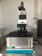
Raman spectroscopy set-up. Double spectromator is a Spex 1404 focal length 85 cm with a pair of 1800 mm-1 diffraction gratings. The excitation lasers are dual 457nm and 515nm (both 50mW) from cobolt, a green (532 nm , 150 mW) laser from Elforlight , a yellow(561nm, 50 mW) and red (660 nm, 100mW) from Cobolt, and of no-use red (785 nm, 80 mW) from Crsytalaser. CCD is a Python 100BR from Princeton Insturuments (liquid-N2 cooled, 1350 x 100 pixels, back illumination). |
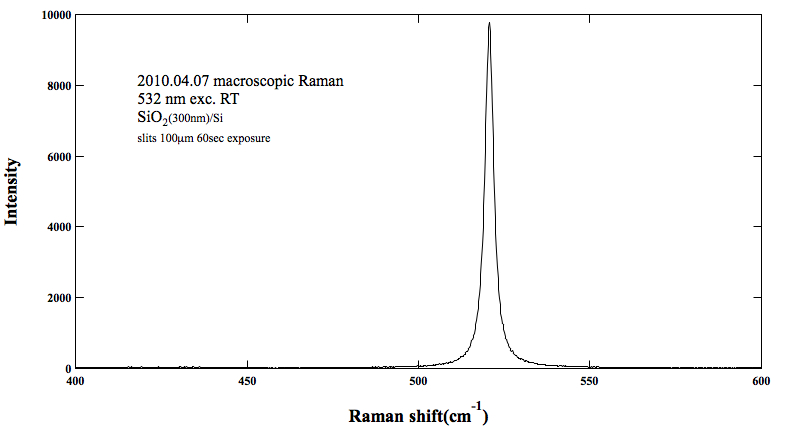
For setting up this macro Raman system, several optical parts and
filters had to be prepared. Prof. Harima and Prof. Gu supplied me
several optics hardwares. The financial supports from Only-One project
and University President's decretion budjet are appreciated. Spectrum
below is the very first Raman signal from Si substrate.
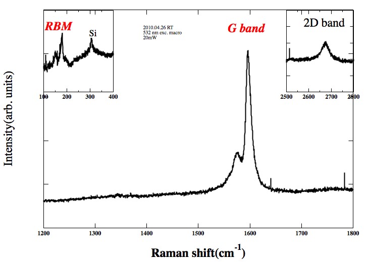
Next two figures are Raman signals from single wall cabon nanotubes.
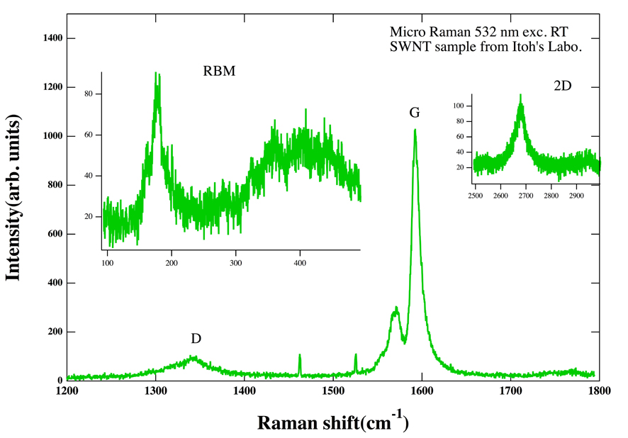
Above figure shows microscopic Raman singal of SWNTs (supplied by Prof. Itoh.)
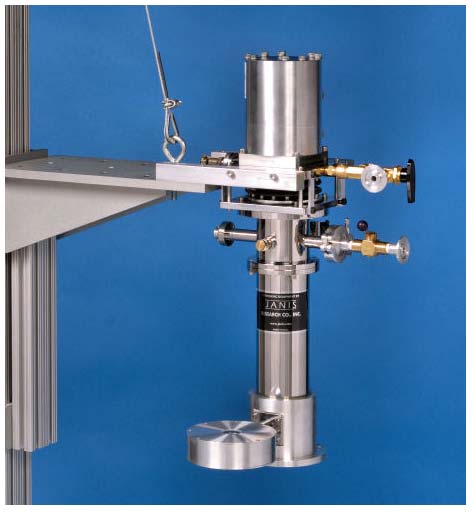
The cryostat below has been equipped with our micro Raman set-up in this March.
Field-emission SEM(JEOL) JSM-7610F and Raman/AFM/SNOM(WITek) Alpha-RAS+ have just added to our research Group.